Serving the Lowcountry and Coastal Empire of Georgia and South Carolina.
Both our Adult and Pediatric Urgent Care Clinics in Savannah will be closed on Sunday, April 20th in observance of Easter Sunday. We will resume normal operating hours on Monday, April 21st.
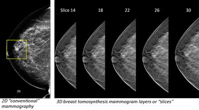
 Breast tomosynthesis uses high-powered computing to convert digital breast images into a stack of very thin layers or “slices”—building what is essentially a “3-dimensional mammogram”.
Breast tomosynthesis is a new technology in the fight against breast cancer. Breast tomosynthesis may be used in conjunction with traditional digital mammography as part of your annual screening mammogram to capture more breast images. Very low X-ray energy is used during the screening examination so your radiation exposure is safely below the American College of Radiology (ACR) guidelines. Using breast tomosynthesis and digital mammography together for screening has been proven to reduce “call-backs”.
Breast tomosynthesis uses high-powered computing to convert digital breast images into a stack of very thin layers or “slices”—building what is essentially a “3-dimensional mammogram”.
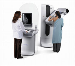
 During the tomosynthesis part of the exam, the X-ray arm sweeps in a slight arc over the breast, taking multiple breast images in just seconds. A computer then produces a 3D image of your breast tissue in one millimeter layers.
Now the radiologist can see breast tissue detail in a way never before possible. Instead of viewing all the complexities of your breast tissue in a flat image, the doctor can examine the tissue a millimeter at a time. Fine details are more clearly visible, no longer hidden by the tissue above and below.